Getting an Accurate Diagnosis
Peripheral neuropathy is notoriously underdiagnosed. The typical patient sees three or more doctors over two years before receiving a confirmed diagnosis. Understanding the diagnostic process helps you advocate for thorough evaluation - and reach answers faster.
Step by Step
The Diagnostic Journey
A complete neuropathy workup follows a logical progression - from clinical exam to electrophysiology to specialized lab testing. Most patients don't need every test; your neurologist will narrow the pathway based on findings at each stage.
Step 1 - First Visit
Initial Medical History & Symptom Review
Your doctor documents when symptoms began, how they've progressed, and which body parts are affected. They'll ask about diabetes, alcohol use, toxic exposures, family history, and all medications - including over-the-counter drugs and supplements. Vitamin B6 toxicity from supplements is a common overlooked cause.
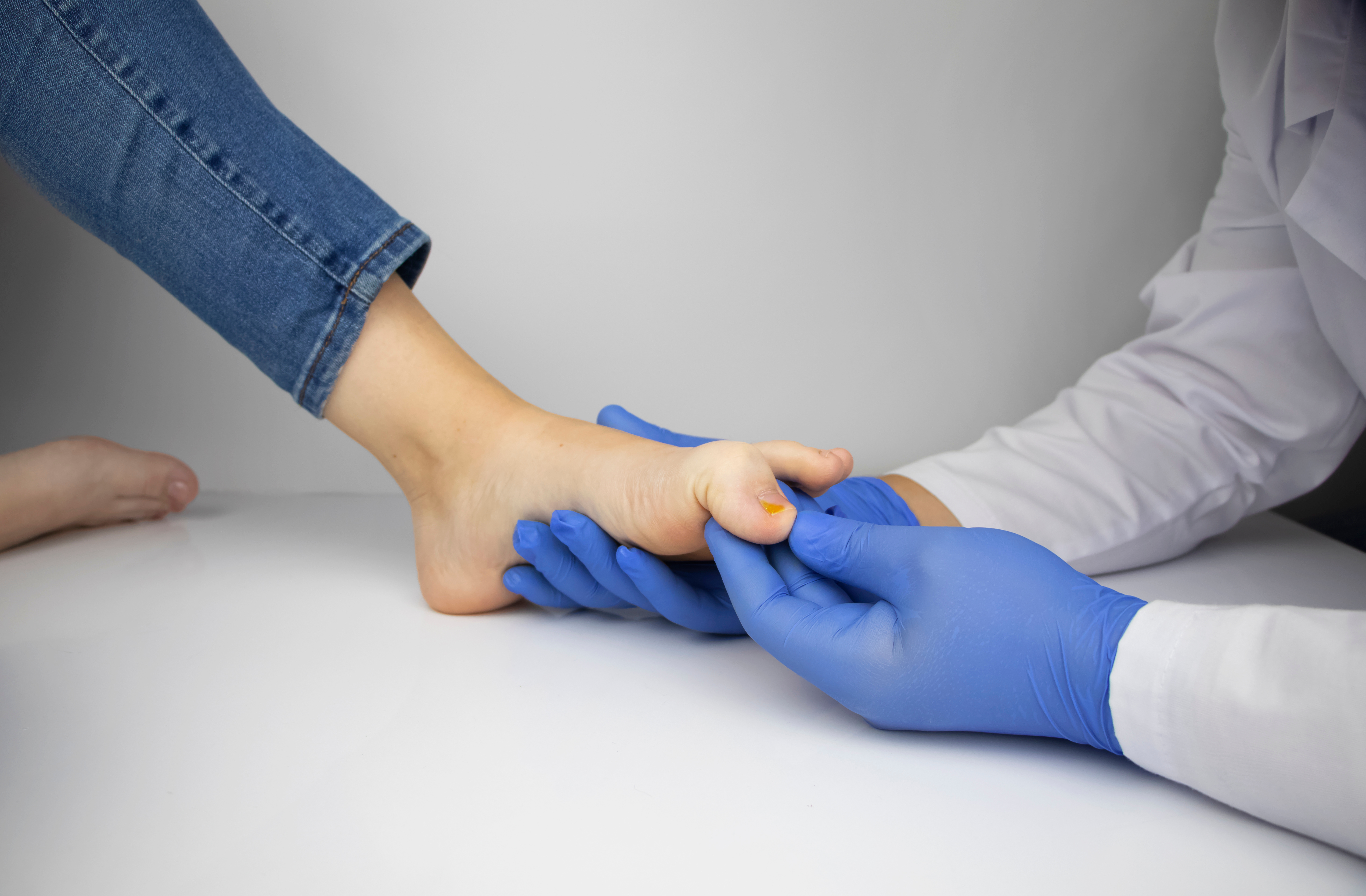
Step 2 - Physical Exam
Neurological Examination
A hands-on assessment testing sensation (light touch, pinprick, vibration, temperature), reflexes at the ankle and knee, muscle strength, and coordination. The physician may use a tuning fork at the big toe to assess large-fiber function, and a monofilament on the sole to screen for protective sensation loss - a critical diabetes-related risk factor.
Step 3 - Electrophysiology
Nerve Conduction Study (NCS)
Small electrodes on the skin deliver brief electrical pulses while sensors measure how fast and how strongly signals travel through specific nerves. NCS is the most objective test for large-fiber neuropathy - it measures conduction velocity and amplitude in motor and sensory nerves. Slowed velocity suggests demyelination; reduced amplitude suggests axonal loss. NCS cannot assess small fibers (C-fibers, A-delta).
Step 4 - Electrophysiology
Electromyography (EMG)
A thin needle electrode inserted into specific muscles records electrical activity at rest and during contraction. EMG reveals whether muscle abnormalities stem from nerve damage (neuropathy), the nerve root (radiculopathy), or the muscle itself (myopathy). It is typically performed alongside NCS in the same session by the same technician or neurologist.
Step 5 - Laboratory
Comprehensive Blood Work
Blood tests identify the most common reversible causes: diabetes, B12 deficiency, thyroid disease, kidney failure, inflammatory disorders, and paraproteinemia. The specific panel ordered depends on your clinical presentation. See the full panel breakdown in the section below.
Step 6 - If Needed
Specialized & Advanced Testing
When initial workup is inconclusive or small-fiber neuropathy is suspected, advanced testing provides critical additional information. Skin punch biopsy - quantifying intraepidermal nerve fiber density - is the gold standard for confirming small-fiber neuropathy, which NCS cannot detect. MRI rules out structural causes; lumbar puncture is reserved for suspected inflammatory or infectious causes.
Laboratory Workup
Standard Blood Test Panel
A systematic blood panel catches the most common and treatable causes. Most neurologists order a core set for every patient, then add targeted tests based on findings and risk factors.
| Test | What It Detects | Notes | Priority |
|---|---|---|---|
| Fasting glucose | Diabetes / prediabetes | Most common reversible cause; ≥126 mg/dL = diabetes | Key Test |
| HbA1c | 3-month blood sugar average | ≥6.5% = diabetes; 5.7-6.4% = prediabetes (also causes neuropathy) | Key Test |
| Vitamin B12 | B12 deficiency neuropathy | Levels 200-400 pg/mL are borderline; MMA and homocysteine may be needed | Key Test |
| Folate (B9) | Folate deficiency | Deficiency can mimic B12 neuropathy; less common but important to exclude | Routine |
| TSH (thyroid) | Hypothyroidism | Hypothyroidism causes length-dependent neuropathy; often overlooked | Key Test |
| CBC with differential | Anemia, infection, blood disorders | Macrocytic anemia (large red cells) suggests B12/folate deficiency | Routine |
| CMP (metabolic panel) | Kidney & liver function | Uremic neuropathy from kidney failure; hepatic neuropathy from liver disease | Routine |
| ESR / CRP | Systemic inflammation | Elevated in vasculitis, inflammatory neuropathies, connective tissue disease | Routine |
| ANA panel | Autoimmune disorders | Screens for lupus, Sjögren's syndrome, rheumatoid arthritis - all cause neuropathy | If Indicated |
| SPEP / SPIP | Paraprotein (M-protein) | MGUS-associated neuropathy is common in older adults; can be treatable | If Indicated |
| Anti-ganglioside antibodies | Immune-mediated neuropathy | Anti-MAG, anti-GM1, anti-GQ1b; identifies CIDP, MMN, and Guillain-Barré variants | If Indicated |
| Vitamin B6 (pyridoxine) | B6 toxicity | Often ordered last but critically important; many patients take high-dose B6 supplements | If Indicated |
Advanced Testing
Specialized Diagnostic Tests
Skin Punch Biopsy
Small-fiber neuropathy gold standard
A 3mm punch biopsy removes a small cylinder of skin (typically from the calf and thigh), which is then stained and analyzed under a microscope to count intraepidermal nerve fiber density (IENFD). Values below age- and sex-matched norms confirm small-fiber neuropathy.
This is critical because NCS is entirely normal in pure small-fiber neuropathy - patients with severe burning pain and NCS showing "no abnormality" often have SFN confirmed on biopsy. Procedure takes 15 minutes; mild local anesthetic. Results in 2-4 weeks.
MRI (Spine & Nerve)
Structural and inflammatory causes
MRI of the lumbar or cervical spine rules out disc herniation, spinal stenosis, or cord compression mimicking peripheral neuropathy. MRI neurography (MRN) directly images peripheral nerves, detecting nerve compression, hypertrophy in CIDP, or perineural infiltration in malignancy.
MRI is not a routine first-line neuropathy test but becomes essential when symptoms are asymmetric, involve the upper limbs prominently, or when compressive radiculopathy needs to be excluded before planning treatment.
Lumbar Puncture (Spinal Tap)
Inflammatory & infectious neuropathies
CSF analysis showing elevated protein with normal white cell count (cytoalbuminous dissociation) is a diagnostic hallmark of Guillain-Barré syndrome and CIDP. It can also detect Lyme disease, CMV, and other infectious neuropathies.
Reserved for cases where immune-mediated neuropathy is strongly suspected - particularly rapidly progressive weakness, prior viral illness, or when IVIG/plasmapheresis treatment is being considered. Most chronic neuropathy patients will never need this test.

What to Bring to Your Appointment
Thorough preparation shortens the diagnostic timeline significantly. Neurologists make faster and more accurate assessments when patients arrive with organized documentation.
- Complete medication list - prescription, OTC, supplements with doses and start dates
- Symptom timeline - when did symptoms start, what body parts, how they've changed
- All prior bloodwork - glucose, HbA1c, B12 results from the past 2 years
- Family history - hereditary neuropathy (CMT), diabetes, autoimmune disease in relatives
- Occupational & toxic exposures - solvents, heavy metals, pesticides, alcohol history
- Shoe and sock photos or samples - abnormal wear patterns reveal gait and weakness
- Pain diary - worst times of day, triggers (heat, walking, rest), quality (burning vs stabbing)
- A support person - neurological appointments cover a lot; a second listener helps retention
Financial Planning
What Diagnostic Testing Costs
Costs vary widely by region, facility, and insurance plan. These figures represent typical ranges in the United States as of 2025. Always verify prior authorization requirements with your insurer before scheduling electrophysiology studies.
With Insurance (after deductible)
Without Insurance (self-pay)
Tip: Negotiated self-pay rates through direct lab companies (e.g., Quest, LabCorp) can reduce blood panel costs to $50-$150 for a comprehensive neuropathy workup. Freestanding imaging centers often charge 40-60% less than hospital-based MRI. Always ask for the cash-pay rate before paying the sticker price.